
A team of scientists from GANIL developed a scintillation dosimeter named SCICOPRO (SCIntillation for the COntrôle des irradiations en PROtonthérapie) in collaboration with the Caen Corpuscular Physics Laboratory and the François Baclesse Cancer Center.
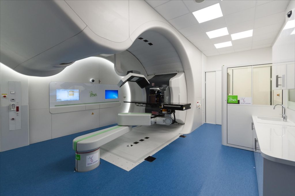
This center operates a proton therapy device called Proteus ONE (IBA company) equipped with ‘pencil beam scanning,’ which delivers irradiation using multiple extremely fine beams that scan the tumor volume. Before irradiating a patient, medical physicists perform a quality control of the treatment by measuring the dose profile in depth on a water phantom. This step is time-consuming as it requires the use of several instruments.
The study conducted here led to the development of a unique system for performing these quality control measurements and reducing the time dedicated to this task.
An evaluation with known beams demonstrated that SCICOPRO was capable of measuring proton beams with an accuracy of approximately 580 µm in position, 180 keV in energy, and 3% in intensity. Measurements of the system for an actual treatment plan with several thousand beams delivered at 1 kHz also showed that this new scintillator-based device verifies the beams to be delivered to the patient. It is even sensitive to errors in the orientation of the treatment table, as small as 1°. SCICOPRO’s performance is very promising and meets the needs of clinical practice.
The results were published in the journal Medical Physics
Before this new device can be used in routine clinical practice, a larger prototype is under development, and the 3D dose distribution measurement is the subject of a PhD thesis.
How SCICOPRO Works
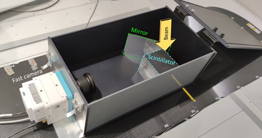
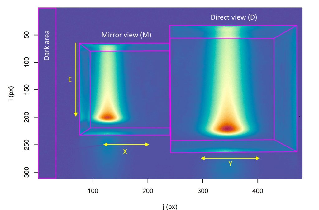
The developed system (Figure 1) is based on the use of a 10 x 10 x 10 cm³ plastic scintillator that emits visible light (scintillation) when irradiated. An ultra-fast CMOS camera records the scintillation produced by each elementary beam delivered in proton therapy. A mirror positioned at 45° relative to the cube allows two views of the scintillator to be acquired (Figure 2).
For quality control of the treatment, it is placed on the examination table instead of the patient. Depending on the treatment, the number of beams delivered can range from a few hundred to several thousand, with scanning durations of a few minutes.
The pulse images are then summed to reconstruct the beam images and analyzed to determine their characteristics. The light distributions measured are representative of the energy deposition of the protons. The position of the light distribution in the image allows the position of the beam in the treatment room’s reference frame to be identified, the depth of the distribution is determined by the beam’s energy, and the light intensity corresponds to its intensity.
Performance Evaluation
Initially, the performance of SCICOPRO was evaluated using beams with known characteristics. This evaluation demonstrated that SCICOPRO could measure all beam characteristics of a treatment plan (from several hundred to several thousand) in a single acquisition, with an accuracy of approximately 580 µm in position, 180 keV in energy, and 3% in intensity. These performances are very promising and meet the needs of clinical practice.
Verification of Proton Therapy Treatment Plans
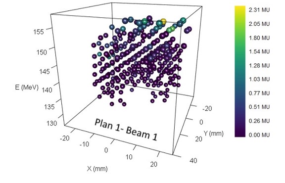
After validating its performance, SCICOPRO was used to verify the agreement between the characteristics of the beams delivered by the irradiation system and the planned characteristics prior to treatment. To do this, various patient treatment plans were studied. Figure 3 presents an example of measuring beam characteristics using SCICOPRO under nominal irradiation conditions. For clarity, only the measured values are shown, but they correspond to the planned values.
Measurements were also made by introducing errors in the positioning of the treatment table to test SCICOPRO’s ability to detect them. These tests showed that it is possible to detect orientation errors as small as 1°.
Impact in proton therapy
he use of proton beams in radiotherapy offers advantages over photon therapy because charged particles have a limited range, which helps to better spare healthy tissues in many indications. In addition to this characteristic, proton therapy treatments are increasingly delivered using fine beams that sweep the tumor volume, providing better dose conformity.
However, the number of fine beams required for a treatment can reach several thousand, and their delivery is divided into several non-consecutive fractions (or pulses) to minimize uncertainty about the intensity delivered during treatment. The pulses are delivered at 1 kHz, meaning one pulse every millisecond.
Verifying the planned beam characteristics (X and Y positions, energy E, and intensity I) for a treatment is therefore a challenging and time-consuming task in the clinic, especially since dosimetry tools are fewer in proton therapy than in photon therapy. Most commercial dosimeters have limitations such as reduced spatial and temporal resolution, and only allow measurements in 1 or 2 dimensions. Verifying all characteristics therefore requires multiple measurements with different detectors.
To overcome these limitations and provide a system that reduces the time spent on quality control in the clinic, scintillation detectors are highly promising. They offer excellent spatial resolution (< 1 mm) and have the potential to enable 3D measurements.
Advances in camera speed also present an opportunity in this field, enabling the verification of treatments with fine beams. The prototype’s size limits the size of the irradiated fields to 6 x 6 cm². However, a larger prototype based on a 25 x 25 x 25 cm³ scintillator is also under study. Furthermore, the measurements presented here are limited to verifying the characteristics of fine beams. The dosimeter could allow for more comprehensive treatment control, including measuring the 3D dose distribution during treatment. This is currently the subject of a PhD thesis.
Contact Ganil: Anne-Marie FRELIN