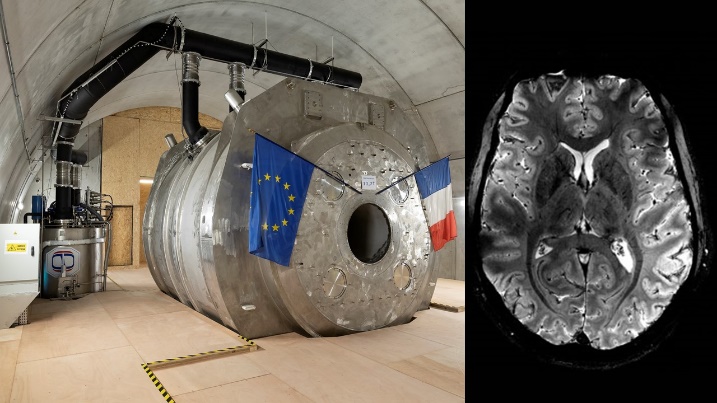
The Iseult project has unveiled the first human brain images obtained using a 11.7 teslas MRI, after almost 25 years of work. This world first was made possible thanks to the commitment of over 200 CEA employees, who believed in this extremely ambitious project from the very beginning
In the early 2000s, a Franco-German project was launched to develop ultra-high resolution imaging. One of the objectives was to build an imager whose key component was a superconducting magnet reaching 11.7 Tesla with a 900 mm aperture, but there was at this time no MRI manufacturer ready to embark on this crazy adventure alone. Based on its strong expertise in superconducting magnets acquired over the past 40 years, in particular for high energy physics and particle physics (Cern) as well as for fusion (Tore Supra, ITER), CEA decided to take up the challenge. After only a few years of design work, CEA proposed in 2006 an initial design using several innovative technological solutions. After exhaustive tests to validate all of them with several prototypes, the final fabrication started in in 2010. It took 7 years for the CEA and Alstom (now General Electric) teams to finalize the construction of this outstanding magnet, a colossus weighing 132 tons, 5 me in length and 5 meters in diameter. The magnet winding is made of 182 km of superconducting wires cooled to -271.35°C by 7,500 liters of superfluid helium.
.
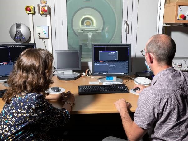
The commissioning of the 11.7 T Iseult MRI in 2021 crowned almost 20 years of AOC research and development. In an article published in the journal Magnetic Resonance Materials in Physics, Biology and Medicine, Nicolas Boulant and Lionel Quettier, Iseult project leaders for the CEA's Joliot and Irfu Institutes, review the details of this commissioning.
Within the framework of a collaborative project between the DES/DDSD and the DRF/IRFU, a feasibility study of muography potential for the auscultation of nuclear reactors was initiated in 2017. After an initial evaluation phase carried out by IRFU using numerical modelling, first data were taken on the G2 reactor block, located at CEA Marcoule and shut down in the early 1980s, from February 2020. These measurements demonstrated the potential of the technique, identifying differences between the current structure of the G2 reactor and the 3D model created from the original plans of the installation. These initial results demonstrate the interest of using muography in the clean-up and dismantling of nuclear facilities, one of the CEA's priorities nowadays. For the next phase of the project, a 3D tomography of the reactor is envisaged by combining images taken from different positions. It could be the first 3D image of the interior of a reactor at dismantling phase without using any artificial ionizing radiation. This will provide a new inspection tool to the existing palette.
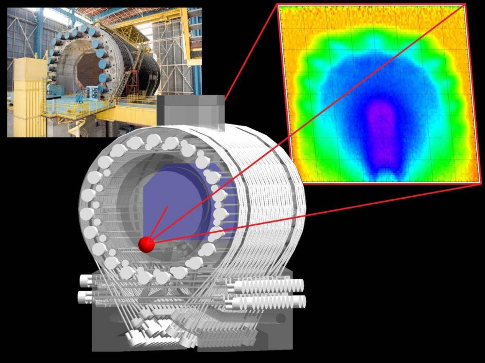
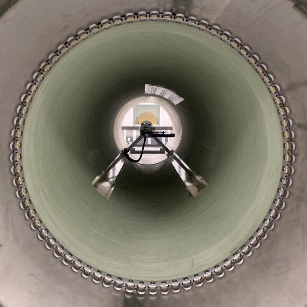
In order for the images produced by the future MRI to be free of distortions or artifacts, the magnetic field generated by the Iseult magnet must be homogeneous to 0.5 PPM (parts per million) around the patient's brain. To meet this challenging specification, it was necessary to provision means of "shimming" the field, i.e. of correcting all the small defects that would inevitably arise from the manufacturing process. 5904 pieces of shim (small iron platelets) were screwed onto rails and installed inside the magnet tunnel. This first configuration was tested on Thursday, July 9, 2020 by mapping its effect on the magnetic field of Iseult at 3 T. The results are very encouraging as this first shimming iteration allowed to increase the homogeneity of the field in the useful zone from 138.8 to 3.2 PPM (value extrapolated to 11.72 T from magnetic measurements at 3 T).
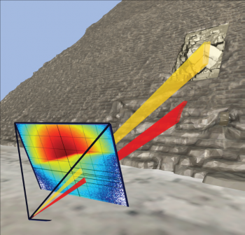
In its most common version, muon imaging is intrinsically a 2D technique: the resulting density map is indeed integrated along the observation direction. However, a 3D map can be obtained by combining several projections, like for medical imaging. But in the muon case, the number of projections is dramatically reduced because of the required acquisition time. A 3D algorithm has been recently developed using the Irfu TomoMu setup, within a collaboration between Florence University and Irfu. The 3D structure of the test object has been reconstructed from only 3 points of view, thanks to the high precision of the instrument. This technique will soon be extended to more applications, from reactor dismantling to civil engineering or mining exploration.
![]() PHoCEA DSM 2024 - Tous droits réservés - Mentions légales - Ce site utilise Twitter Bootstrap
PHoCEA DSM 2024 - Tous droits réservés - Mentions légales - Ce site utilise Twitter Bootstrap
Dernière mise à jour : Nov 19th, 2024

