
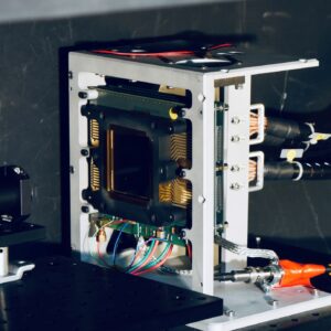
Ionizing radiation detectors with very high temporal resolution are key elements not only in particle physics, but also in medical imaging. Capitalizing on its expertise in gamma radiation detection (scintillating crystal calorimeters) and in fast analog and digital electronics, the Group has been developing detectors for functional PET medical imaging since 2009. A PET image is obtained by injecting into the patient’s body a biochemical and radioactive tracer involved in the biological process to be visualized. The tracer emits a positron which annihilates with a nearby electron, producing two 511 keV photons. Detection of the photons gives access to the positron’s emission position. PET imaging enables us to see the body’s biochemistry at work, in its rarest processes.
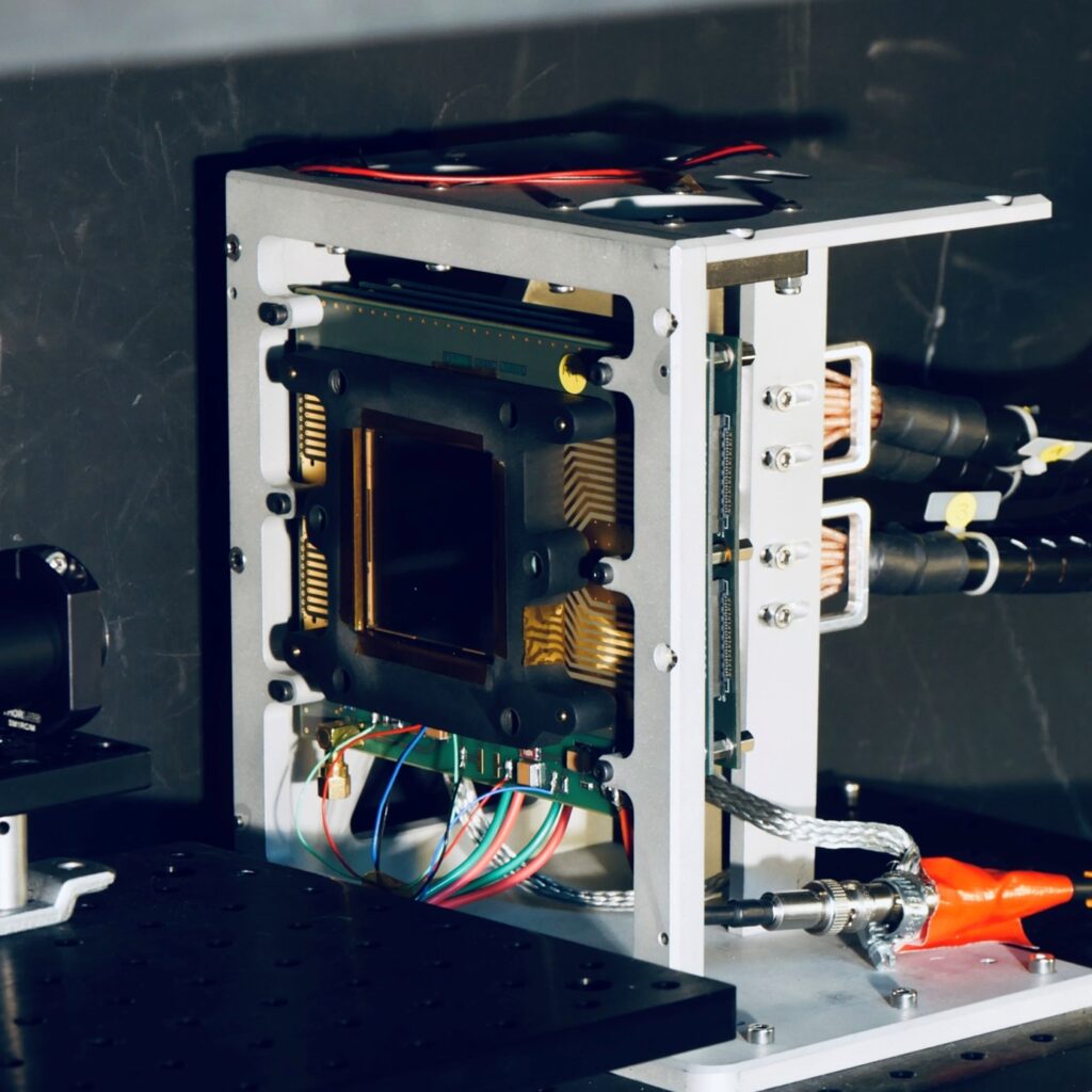
The group’s aim is to develop innovative technologies that can bring decisive advances over commercial imagers, in particular by improving temporal resolution and thus image contrast. To this end, the group has been associated with UMR BioMaps at Orsay since 2020. The CLEARMIND project, coordinated by DPhP, aims to develop detectors with high temporal resolution (
Contact: Dominique Yvon